Conditions
Unilateral Biportal Endoscopic (UBE) Surgery of the Lumbar Spine


Unilateral Biportal Endoscopic (UBE) Surgery of the Lumbar Spine
Five (less often four or six) lumbar vertebrae (L1 to L5) and the sacrum (S1) form the lower part of the spine, which bears most of the body's weight and provides spinal mobility. At this level, the spinal canal contains the termination of the spinal cord and the nerve roots arising from it (the so-called cauda equina), which innervate the legs, pelvic organs, and bowel / lower pelvic organs, such as the bladder and genitals.
Intervertebral discs absorb physical load and allow the vertebrae to move. Each intervertebral disc has a strong outer (fibrous) ring and a soft inner core. Problems with intervertebral discs can be caused by sudden overload, such as lifting heavy objects, trauma, or prolonged everyday wear and tear.
When the spine is healthy, the vertebrae and intervertebral discs of the lumbar spine allow the following movements:
• bending forward / backward (flexion and extension);
• lateral bending (side bending);
• turning to the right and left (rotation).
What are degenerative changes in the lumbar spine?
Degenerative changes in the lumbar spine are a normal part of the aging process of the spine. These changes are not always associated with pain or a reduction in quality of life. Study data show that degenerative changes in the intervertebral discs on imaging (MRI) are observed in approximately 37% of people at age 20, but already in approximately 96% of people at age 80, with the greatest increase occurring up to age 50.
The onset and progression of degenerative changes in the intervertebral discs can be accelerated by, for example, heavy physical work and lifting heavy objects, prolonged sitting (office or driving work), excess body weight, smoking, and prolonged exposure to vibration. These factors can cause damage to the intervertebral discs, which may lead to compression of one or more nerve roots and pain / sensory disturbances in one or both legs.
Disc degeneration can lead to the following consequences:
- Disc herniation, which can cause compression of a nerve root and pain radiating into the leg, tingling, or weakness in the leg (radiculopathy).
- Narrowing of the spinal canal (spinal stenosis), which can cause pain and weakness in the legs when walking or standing (neurogenic claudication) that eases when sitting down or bending forward. In rare, severe cases, a large intervertebral disc herniation and pronounced stenosis can cause cauda equina syndrome with bowel and/or bladder dysfunction - a condition that requires urgent surgical intervention.
- Narrowing of the intervertebral foramen or the lateral segment of the spinal canal, in which the symptoms are similar to those of disc herniation.
What is biportal endoscopic spine surgery (UBE)?
Unilateral Biportal Endoscopy (UBE) is a minimally invasive spine surgery method in which the surgeon inserts two instruments through two separate skin incisions, each approximately 8 mm, on one side of the spine. Through one incision (the viewing portal), an endoscope with a high-resolution camera is inserted, providing a magnified, well-illuminated image of the surgical field, while through the other opening (the working portal), surgical instruments are inserted (a high-speed burr, a radiofrequency probe, a soft-tissue resection instrument or shaver, Kerrison rongeurs, and others). A continuous flow of sterile saline solution through the surgical field ensures clear visibility and helps control bleeding.
Unlike uniportal endoscopic surgery, in which the endoscope and instruments are inserted through the same channel, in the UBE method the two portals are separate and can move independently of each other, which allows the use of standard microsurgical instruments and gives the surgeon greater freedom of manoeuvre.
How is UBE surgery performed?
The surgery is usually performed under general anesthesia, with the patient lying face down. Using X-ray (fluoroscopy) guidance, the surgical spinal level is precisely identified and the locations of both portals are marked. Two incisions of approximately 8 mm are made through the skin, subcutaneous tissue, and muscle fascia. Through the working portal, using a burr and other instruments, part of the vertebral lamina and the ligamentum flavum are partially removed to access the spinal canal and the nerve root. Depending on the diagnosis, the damaged disc fragment is removed (discectomy) or the narrowed spinal canal is widened (decompression). After surgery, the skin incisions are closed with cosmetic (intracutaneous) sutures or skin glue, and the patient can go home on the evening of surgery or the following day.
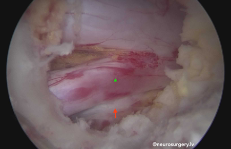
Image: Access to a right-sided L5/S1 disc herniation (red arrow) compressing the right S1 nerve root (green star), performed using the biportal spinal endoscopy method.
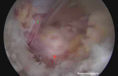
Image: Access to a right-sided L5/S1 disc herniation, performed using the biportal spinal endoscopy method. The intervertebral disc herniation (red arrow) after mobilization and retraction of the right S1 nerve root (green star) with a nerve root retractor.
In which cases is endoscopic spine surgery recommended?
- Intervertebral disc herniation with symptoms of radiculopathy - pain, tingling, or weakness in the leg.
- Foraminal (intervertebral foramen) or lateral recess spinal canal stenosis (narrowing).
- Central spinal canal stenosis with symptoms of neurogenic claudication (reduced walking distance).
- The patient has undergone at least six weeks of unsuccessful non-surgical (conservative) treatment, except in cases requiring urgent surgery (for example, severe pain, pronounced or progressive leg weakness, or cauda equina syndrome).
In which patients is endoscopic spine surgery not recommended? What are the contraindications?
If any of the following conditions is diagnosed, UBE surgery may not be suitable or may be contraindicated:
- pronounced segmental instability of the vertebrae requiring spinal stabilization (fusion) surgery;
- vertebral fracture, tumor, or active infection in the segment of the spine to be operated on;
- uncorrectable coagulation disorders or anticoagulant therapy that cannot be temporarily discontinued;
- extensive changes affecting multiple levels, for which microsurgical or open surgery is more suitable;
- serious comorbidities that preclude surgery under general anesthesia;
- pregnancy;
- pronounced obesity that significantly hinders access and visualization;
- the patient's inability to remain lying face down for the required duration of the surgery;
- local skin or soft-tissue infection at the intended surgical site.
What complications are possible with endoscopic spine surgery?
Study data show that unilateral biportal endoscopy (UBE) surgeries are generally safe - in studies, the overall complication rate in the endoscopic spine surgery group (~5%) was lower than in the traditional microscopic surgery group (~17%). However, as with any other spine surgery, similar complications can also occur with endoscopic spine surgery:
- injury to the dura mater with cerebrospinal fluid (CSF) leakage (in approximately 2–4% of cases);
- post-operative epidural hematoma;
- temporary sensory disturbances or unpleasant sensations (dysesthesia) in the leg, which may be caused by the pressure of the irrigation fluid or irritation of the nerve root during surgery;
- possible development of instability in the operated segment - the future need for spinal fixation surgery;
- incomplete removal of the disc fragment or incomplete decompression, which may require a repeat operation;
- recurrence of disc herniation (repeat herniation) at the same level;
- error in determining the surgical level;
- superficial or deep wound infection (less common than with open surgery);
- injury to a nerve root/roots: a sensation of weakness or numbness in the leg;
- dysfunction of the lower pelvic organs - disturbances of bowel or bladder function, sexual dysfunction;
- injury to blood vessels;
- rarely - increased pressure in the epidural space or spinal canal due to the irrigation fluid;
- reaction to medications or anesthetic agents during or after surgery;
- blood clot (thrombosis) in the bloodstream or pulmonary embolism.
What is the difference between UBE surgery and traditional microsurgical surgery?
Instead of endoscopic spine surgery, patients often undergo traditional microsurgical spine surgery (for example, microdiscectomy or spinal canal decompression), using an operating microscope and a larger skin/muscle incision to access the damaged disc or narrowed spinal canal. The goal of both methods is the same - to relieve the compressed nerve roots or spinal cord and eliminate the cause of the pain and functional disturbances.
The main advantage of UBE surgery is its less invasive approach - small skin incisions, less damage to muscle and bone tissue, and - thanks to the endoscope - better visualization and magnification of the surgical field.
As a result, the advantage of endoscopic spine surgery is usually less post-operative pain, less blood loss and risk of infection, better healing of surgical wounds, and consequently - faster recovery.
Both traditional microsurgical surgery and endoscopic spine surgery:
- relieve compression of the nerve roots and spinal cord;
- eliminate the pressure on nerve structures caused by the damaged disc fragment or narrowing;
- can be performed at one or more spinal levels.
Advantages of endoscopic spine surgery, compared with open microsurgical surgery:
- better visualization and magnification of the surgical field;
- minimal scarring process;
- lower risk of infection;
- less post-operative pain;
- faster recovery process.
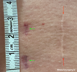
Image: Green arrows: skin scars 1 month after L4/5 endoscopic discectomy surgery on the left side. Red arrows: skin scar 11 years after L4/5 microdiscectomy surgery on the right side.